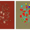
. The data cube can be divided into clusters (by hierarchical cluster analysis) and thus converted into a compositional map (bottom panel). It reveals the polymer components (grey, blue and red areas), as well as the interfaces between them (green areas) that partially exhibit anomalies that are explained by chemical interaction (purple areas). Copyright: CIC nanoGUNE.")
An ultimate goal in materials science, biomedicine or nanotechnology is the non-invasive compositional mapping of materials with nanometre-scale spatial resolution. A variety of high-resolution imaging techniques exist (for example, electron or scanning probe microscopies), however, they cannot meet the increasing demand in research, development and industry of being non-invasive while offering the highest chemical sensitivity.
Nanoscale chemical analysis has become possible with nano-FT-IR spectroscopy, that combines scattering-type scanning near-field optical microscopy (s-SNOM) and Fourier transform infrared (FT-IR) spectroscopy. By illuminating the metallised tip of an atomic force microscope (AFM) with a broadband infrared laser or a synchrotron and analysing the backscattered light with a specially designed FT spectrometer, local IR spectroscopy with a spatial resolution of <20 nm has been demonstrated. However, only point spectra or spectroscopic line scans comprising no more than a few tens of nano-FTIR spectra could be achieved on organic samples, owing to the long acquisition times.
Now, researchers from CIC nanoGUNE (San Sebastian, Spain), Ikerbasque (Bilbao, Spain), Cidetec (San Sebastian, Spain) and the Robert Koch-Institut (Berlin, Germany) have developed hyperspectral infrared nanoimaging. The technique allows for recording two-dimensional arrays of several thousand nano-FTIR spectra (hyperspectral data cubes) in a few hours and with a spatial resolution and precision better than 30 nm.
“The excellent data quality allows for extracting nanoscale-resolved chemical and structural information with the help of statistical techniques (multivariate data analysis) that use the complete spectroscopic information available at each pixel”, says Iban Amenabar, first author of the work published in Nature Communications. Even without any previous information about the sample and its components, pixels with similar infrared spectra can be grouped automatically with the help of hierarchical cluster analysis. By imaging and analysis of a three-component polymer blend, the researchers obtained nanoscale chemical maps that not only reveal the spatial distribution of the individual components but also spectral anomalies that were explained by local chemical interaction. The researcher also demonstrated in situ hyperspectral infrared nanoimaging of native melanin in human hair.
For their experiments, the researchers used the commercial nano-FT-IR system from Neaspec GmbH including a mid-infrared laser continuum that covers the spectral range from 1000 cm–1 to 1900 cm–1. Multivariate analysis of the hyperspectral data was performed with the software tool CytoSpec, which was developed by co-author Peter Lasch.
“With the rapid development of high-performance mid-infrared lasers and by applying advanced noise reduction strategies, we envision high-quality hyperspectral infrared nanoimaging in few minutes”, concludes Rainer Hillenbrand who led the work. “We see a large application potential in various fields of science and technology, including the chemical mapping of polymer composites, pharmaceutical products, organic and inorganic nanocomposite materials or biomedical tissue imaging”, he adds.

; 3,5-tetrabromobisphenol A, TBBPA (yellow samples); Pentabromophenyl ether, Deca-BDE (green samples); and reference, REF (blue samples).")
. Oslo kommunes kunstsamling (City of Oslo Art Collection).")


 of an ulcer with delayed healing.")