Cancer continues to be a global health threat, responsible for millions of lives lost each year. Standard cancer treatments like chemotherapy and radiation therapy remain the primary methods for treating neoplasms. However, a small percentage of treated tumour cells, called "therapy-induced senescent" (TIS) cells, exhibit resistance to conventional therapies, leading to tumour quiescence and ultimately, recurrence.
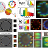
Researchers developed and combined advanced optical microscopy techniques to uncover the biological mechanisms behind the formation of TIS cells in human tumours. One approach involved the use of ultra-short laser pulses, with an incredibly brief duration of just one-millionth of a millionth of a second; this microscopy technique, called “coherent Raman spectroscopy”, allowed the team to identify the chemical constituents of the different biomolecules based on their characteristic mechanical vibrations at high frequency. Another approach was based on “phase holo-tomography”, an advanced imaging method that measures how laser light waves change as they pass through tumour cells; this method allowed the researchers to create three-dimensional reconstructions of the morphology of cells and visualize their inner structures.
The results are very promising. The research group was able to distinguish key features of TIS cells in human tumour cells, shedding light on their early manifestation. These properties include the reorganization of the mitochondrial network, overproduction of lipids, cell flattening, and enlargement. By analyzing a considerable number of cells, researchers established a clear timeline for the development of these distinctive signs.
This result is a clear example of how cutting-edge technologies, multidisciplinary expertise, and strong international collaborations are crucial in addressing the most pressing biological questions, such as the early reaction mechanisms of tumour cells to anticancer therapies.
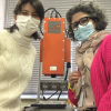
The findings provide important insights into the complex world of TIS in human tumour cells. In our laboratory at Politecnico di Milano, we have developed a new laser microscope that has allowed us to understand the initial stages of this phenomenon. Importantly, this study was conducted without the use of invasive techniques, preserving the natural state of the cells.
The study offers many avenues for the future of cancer research, opening new paths for investigation. The research team envisions broader applications in the development of personalized treatments using patient-derived cancer cells and the potential to refine current screening protocols for oncological therapy. The discoveries made by this research team bring us closer to understanding the complexities of cancer and offer hope for more effective therapies in the future.
The full study can be found at this link: https://doi.org/10.1126/sciadv.adg6231


