Catherine E. Nicholson,a Andrew Beebyb and Richard Gamesonb
aNorthumbria University, Newcastle Upon Tyne NE1 8ST, UK. E-mail: [email protected]; @teampigment; blog: www.durhamgospels.blogspot.co.uk
bDurham University, South Road, Durham DH1 3LE, UK
“Seo nydþearf feala læreð”—Necessity teaches many things.1Durham proverbs, DCL B_II_32 f.43–45
Introduction
Investigation of the pigments used by medieval illuminators is often piecemeal and slow, simply due to the logistics of uniting a medieval manuscript with a spectrometer. Insurance and conservation issues prevent the transport of precious books to scientific facilities for analysis, and the moratorium on sampling results in the fact that only optical spectroscopy (or those using electromagnetic radiation in a non-contact, non-invasive manner) can be used. The techniques of choice are Raman and diffuse reflection [also known as FORS, fibre-optic (visible and near infrared) reflectance spectroscopy] spectroscopies, multi- or hyperspectral imaging and X-ray fluorescence, XRF. Moving bulky equipment to the manuscripts has also been a logistical challenge but recent developments in portable equipment have allowed this area to flourish.
Extreme care must be taken when working with priceless artefacts; by working with conservators, safe exposure limits may be determined. In Raman spectroscopy studies for example, laser power density is the primary concern, with limits of <0.02 mW µm–2 classed as the safe limit to prevent degradation of photosensitive pigments in a confocal microscope.2,3 Often workers are not aware of the conditions they are using, and some commercial systems use far greater power densities to achieve good signal-to-noise ratio, and can rarely adhere to such strict limits. We would encourage anyone considering a purchase for this purpose to be mindful that the technology should not inadvertently damage their artefact.
The necessity for developing better systems has resulted in “Team Pigment” (see http://www.durhamgospels.blogspot.co.uk and @teampigment) working with the instrument manufacturers to develop custom solutions to allow for both portability and sensitivity at such low power densities, and by employing a selection of techniques such as Raman spectroscopy, multispectral imaging and reflection spectroscopy to identify pigments in use on a host of manuscripts.
Moving the mountain
For our initial study on the Northumbrian insular manuscripts of the 7th and 8th centuries,4 we used a Horiba LabRAM HR. This laboratory instrument was moved to the special collections library and was fitted with a re-engineered x,y sample stage to support large manuscripts beneath the microscope head. Neutral density filters allow for the power density to be reduced to safe limits and a 50× Leica LWD (Long Working Distance) objective kept a safe working distance away from the manuscript. This system also had the advantage of multiple lasers to choose from, thus if a particular pigment was fluorescent or absorbing in the region of our standard 633 nm HeNe laser, a 785 nm or 532 nm laser could be used. The utmost care was taken to measure the laser power using a power meter placed at the focal point of the microscope prior to any measurements on manuscripts.
This early work was assisted by the generous provision of two engineers to move and re-align the spectrometer used in this project. Despite expert assistance this move required several days, and muscle power to physically move a laser, optical table and ancillary equipment. It was immediately apparent that whilst this system excelled in performance it was not practicable to move the instrument to other libraries where more work was needed to be done.

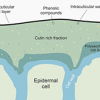
Figure 1. Left to right: manuscript under investigation, overlay of spectrum (orpiment As2S3) from yellow letter fill. Horiba engineers moving spectrometer into medieval dungeon at Palace Green Durham. Portable system in action.
Superheads and supersmall spectrographs
The fixed microscope head of the Horiba LabRAM HR imposed some limitations on which areas of manuscripts could be accessed, hence a Horiba Superhead Raman probe which couples to the laser and spectrometer via fibre optics provided a logical evolution of the equipment. The disadvantage of the Superhead is that the filters are encased within the sealed unit, hence one per laser wavelength is required. However, this Raman probe does allow for coupling to an external laser source, and effectively any spectrograph/detector. Also, because of the fibre coupling of the laser, the spot size of this system is significantly larger than the LabRAM HR so is no longer strictly confocal. The spot is estimated to be 30 µm diameter, giving it a much lower power density at the sample, 0.5 µW µm–2.
Bearing in mind the need for a portable system we chose to assemble a single wavelength mobile Raman spectrometer based upon a low power HeNe laser, the Horiba Superhead and an Ocean Optics QEPro spectrograph/camera as the detection system. This spectrometer, pre-set for operation at 633 nm, has a small footprint, is lightweight and offered good sensitivity. Although limited to a single wavelength, this equipment is readily portable, can be set up in less than half an hour and retains its calibration after a journey.
The system is small enough to fit into a small suitcase and was taken to work in the field for a three-week campaign in the Fitzwilliam Museum in Cambridge. The overall sensitivity of this mobile system was lower than the benchtop system and required longer acquisition times (up to 120 s compared to 20 s on the LabRAM) which slowed progress. This modular system performed admirably and allowed a huge amount of data to be collected from late medieval European manuscripts.
Raman spectroscopy is a powerful technique for the identification of pigments; however, some materials do not readily yield a good spectrum, it can suffer from interference from fluorescent materials and can only analyse a sample on a point-by-point basis. To circumvent this we employ a combination of optical techniques including diffuse reflection and multispectral imaging which reveal the presence of additional pigments, for example carbon- and copper-based inks show low reflectivity in the near infrared spectral region, and cannot be readily detected with the 633 nm portable Raman system at the low power densities considered non-damaging.
Optimisation and testing
Further investment to the “Team Pigment” project has allowed for further refinements to the portable system. We are currently using a deep-cooled, back-illuminated, deep-depletion CCD (charge-coupled device) camera (Andor iDus 416) which offers exceptionally low noise and high-efficiency allowing for reduced acquisition times (typically 20 s) and the possibility of long exposures. Coupled to a spectrograph with a moveable and exchangeable grating (Andor Shamrock 163) this system has the advantage of being able to tune the system to any wavelength, allowing it to be used with a selection of laser wavelengths.
The performance of the Raman probe is an important consideration: several pigments have characteristic bands at low wavenumber, hence they should have as low a low-wavenumber cut-off as possible: systems are now available that can operate at >80 cm–1 for both 633 nm and 532 nm. For example, such a performance allows the identification of mixtures of the lead oxides red lead and massicot, that was previously only possible on a lab-based system.
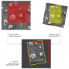
A direct comparison of results collected using the same Raman probe, sample, fibre and incident laser power at the sample of 0.36 mW from these portable systems is shown in Figure 2. In these normalised spectra, a clear improvement in the signal-to-noise ratio (S/N) can be seen with higher performance detection systems. Better S/N could readily be obtained using higher laser powers, but these tests of performance were carried out under similar conditions to those used in the conservation setting.

Figure 2. Comparable results are obtained from different portable detectors using the same collection conditions on liquid sample of toluene, Horiba Superhead and fibre-optic coupling, all using identical acquisition conditions (total power 0.36 mW, 0.5 μW μm–2).
For the conservation community, Raman spectroscopy is not necessarily the silver bullet that permits the reliable identification of pigments, but a useful tool to complement the other techniques already in use. A preliminary study by our group of a selection of common 18th century watercolour pigments revealed that just over half of (24 of the 45) samples provided could be identified using Raman spectroscopy, with this number falling to 20 if just a single laser wavelength was used.
Another vital consideration is the expertise of the user—whilst many conservation scientists excel in a range of fields and are eager to be trained in using this technique, it is one that requires experience and time to perfect. Even after an intensive day-long training course, we find that students and conservators struggle to obtain spectra from test sheets with a thick layer of fresh pigment. This highlights the investment of time required to master the technique, and that it is currently far from a ‘point, click and identify’ solution. Conservator’s time is stretched thinly as it is, and at present we recommend patience whilst the system is refined and optimised for this application rather than making an expensive purchase that may provide little information or, worse, may cause damage to the priceless artefacts they care for.
Conclusion
Whilst Raman spectroscopy is far from its infancy in this field,5,6 it is only now becoming a fully-fledged technique that can be applied in situ to any artefact without causing damage from the light exposure of the incident laser beam. Ongoing development of the equipment by instrument manufacturers (see Table 1) is leading towards new optimised modular spectrometers that can be deployed in the field yet retain or improve upon the performance of their lab-based counterparts at comparable low laser power densities. Development of this portable equipment has allowed relationships between conservation scientists, librarians and historians to grow. “Team Pigment” are pushing the development of the spectrometers in this application and working closely with the manufacturers to make advances in pigment identification in manuscripts.
Table 1. Portable systems currently retailing. Note the authors strongly recommend that the laser power is always measured with a calibrated power meter and the laser power density calculated prior to any measurements on artefacts. (? indicates information not advertised on website.)
Manufacturer | Model | Laser wave-length (nm) | Laser power (min) (mW) | Laser power (max) (mW) |
B&W Tek | i-Raman Plus | 532 | Variable | 50 |
B&W Tek | i-Raman Plus | 785 | Variable | 420 |
B&W Tek | NanoRam | 785 | 30 | 300 |
SciAps | Inspector 500 | 1030 | 300 | 300 |
SciAps | Inspector 300 | 785 | 300 | 300 |
Ocean Optics | IDRaman mini 2.0 | 785 | 50 | 100 |
Rigaku | Progeny | 1064 | ? | ? |
Metrohm | Mira | 785 | ? | 75 |
Metrohm | Mira | 1064 | ? | 400 |
Stellarnet | Various | Custom | Variable | 500 |
BaySpec | Agility | 532 | Variable | 50 |
BaySpec | Agility | 785 | Variable | 499 |
BaySpec | Agility | 1064 | Variable | 499 |
Bruker | Bravo | ? | ? | ? |
Thermo Scientific | TruScan RM | 785 | 225 | 250 |
Renishaw | RA100 Raman analyser | Custom | User specified | User specified |
The final word of caution that we would stress is that laser-based spectroscopic methods such as Raman spectroscopy must be used with the utmost care and consideration such that no damage is done to photosensitive pigments on priceless artefacts, and that conservation limits on light power density always be observed.
Acknowledgements
The authors would like to thank the many libraries, conservation scientists, curators and conservators of the collections we have visited and for allowing us access to the manuscripts in their care.
Notes and references
- As a trained chemist, quoting medieval English was not a prospect I ever imagined, however, spending the past three years as a founding member of “Team Pigment” and carrying out analysis on many manuscripts later, I would like to tip my hat to the original illuminators and scribes to whom I owe my current research interests.
- L. Burgio, R.J.H. Clark and S. Firth, “Raman spectroscopy as a means for the identification of plattnerite (PbO2), of lead pigments and of their degradation products”, Analyst 126, 222 (2001). doi: http://dx.doi.org/10.1039/b008302j
- This is the power density employed in a confocal system, but this does not scale linearly with spot size due to local heating effects.
- Beeby, A.R. Duckworth, R. Gameson, C.E. Nicholson, R.J.H. Clark, B. Meehan and A.W. Parker, “Pigments of the earliest Northumbrian Manuscripts”, Scriptorium 69(1), 33 (2015), ISSN 0036-9772
- B. Guineau, “Analyse non destructive des pigments par microsonde Raman laser; examples de l’azurite et de la malachite”, Stud. Conserv. 29, 35 (1984).
- J. Vezin, “La microsonde Raman Laser: un nouvel instrument d’analyse des pigments dans les enluminures”, Scriptorium 38, 325 (1984)., doi: http://dx.doi.org/10.3406/scrip.1984.1372